
COMPREHENSIVE INTEGRATED PLATFORM FOR TRANSLATIONAL INNOVATION IN OPIOID ABUSE
AnaBios Corporation, in partnership with a team of computational scientists from the Georgia Institute of Technology and the Karlsruhe Institute of Technology in Germany development of a comprehensive integrated platform for translational innovation in opioid abuse. Led by principal investigator Jeffrey Skolnick, Ph.D., from Georgia Tech, and co-investigator Andre Ghetti, Ph.D., Chief Executive Officer of AnaBios, the team has developed an in silico AI-based screening aimed at the identification of novel lead molecules to treat chronic pain without the risk of addiction. The program also integrates validation of the hit compounds in primary human sensory neurons.
This new paradigm was developed and validated with support from the National Center for Advancing Translational Sciences (NCATS) 2020 ASPIRE Reduction-to-Practice Challenge. The NCATS ASPIRE Challenges are part of the of the Helping to End Addiction Long-term Initiative, or NIH HEAL Initiative to speed scientific solutions to the national opioid public health crisis.
End users of the new drug discovery paradigm, can leverage AnaBios’ expertise in human sensory neuron physiological assays and rely on AnaBios’ services to validate the hits from the virtual screening. These experiments will establish the therapeutic potential of the compounds. Alternatively, scientists have the option of downloading the protocol provided below, they can perform the hits validation in their own laboratory if they have access to human sensory neurons.
The multidisciplinary team, whose project focused on the, Virtual screening hits are being validated with AnaBios’ in vitro human primary neuron preparations, and the predictive ability of the combined approach.
Contact us today to validate your virtual hits on human sensory neurons. Please complete the form below and click the “Submit” button. We will respond to you in 24-48 hours.
[ninja_form id=20]
*By Clicking On The “Submit” Button Above, You Are Agreeing To AnaBios’ Privacy Policy.
LIVE CELL IMAGING OF HUMAN SENSORY NEURONS
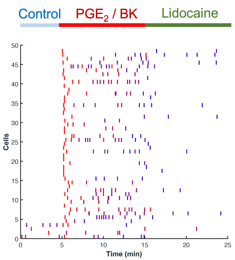
The video below shows live cell imaging of human sensory neurons in culture. Each green circle is the cell body of a neuron. In these experiments, we monitor the spontaneous activation of human neurons by tracking the intracellular calcium level using a fluorescent calcium indicator. Each time a neuron becomes active, it exhibits a brief increase in luminance. During the baseline period, cells are mostly quiescent, but when stimulated with inflammatory agents like PGE2 and bradykinin, the level of activity increase. When a blocker of voltage gated sodium channels is applied, most of the the cells return to quiescence. This kind of data can be analyzed as shown on the right, where a raster plot is used to indicate the activation of each cell with a small vertical bar. From these kind of experiments and analysis, we can determine which molecules selected by artificial intelligence are truly active in human sensory neurons.
NORMALIZED SPONTANEOUS ACTIVITY
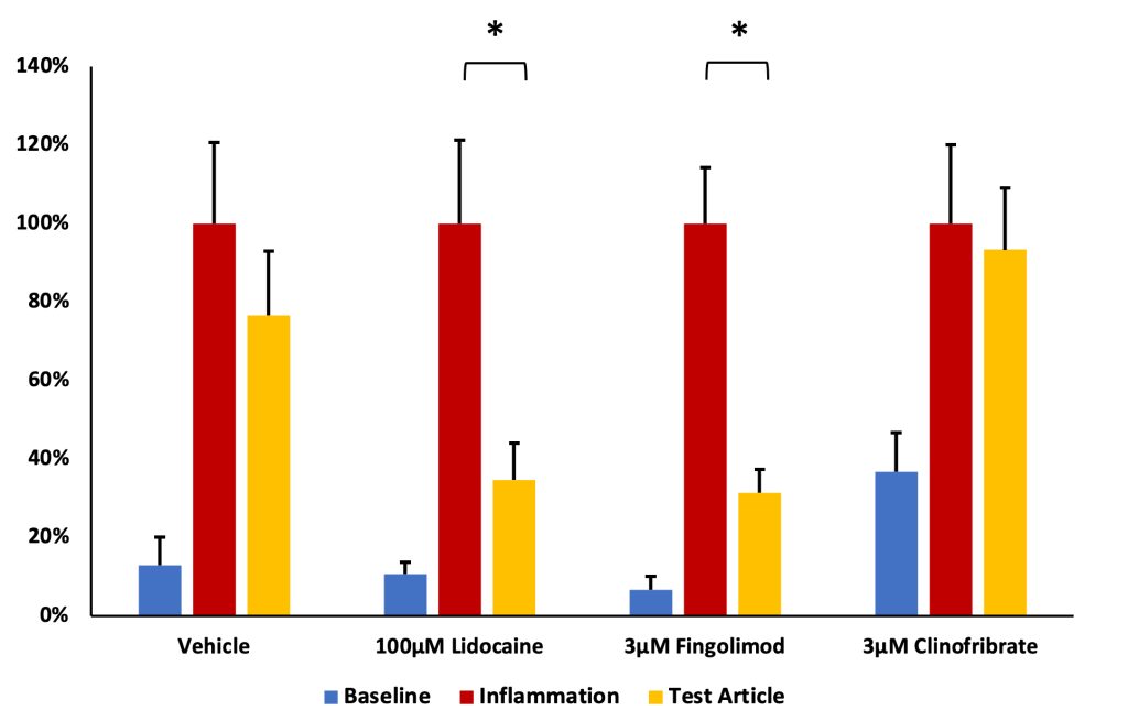
The data from the live cell imaging experiments are summarized in the figure to the left, where spontaneous activity, at baseline (blue bars) and following an inflammatory challenge (red bars) are shown. The reduction of spontaneous activity obtained with the test articles is shown in the yellow bars. The positive control lidocaine and test article fingolimod are both active blockers of neuronal excitability. However, vehicle control or clinofibrate have no effect on neuronal excitability.
PROTOCOL DOWNLOAD
If you would prefer to conduct the study in your laboratory, you may download the protocol for human dorsal root ganglia screening by completing the form below and clicking the “Submit” button.
[ninja_form id=19]
*By Clicking On The “Submit” Button Above, You Are Agreeing To AnaBios’ Privacy Policy.