
HUMAN DORSAL ROOT GANGLIA TISSUE
AnaBios human dorsal root ganglia (DRG) tissue samples are the gold standard biological agent for researchers investigating pain and sensory processing. Our proprietary technology minimizes the ischemic cellular process breakdown, ensuring that our tissue samples maintain high RNA integrity numbers (RIN scores) and superior quality compared to traditional biobanks. Published research demonstrates the viability and functionality of our human DRG tissue samples, which is essential for researchers engaged in target identification and localization. Trust AnaBios to provide you with the highest-quality human DRG tissue samples to accelerate your preclinical drug discovery program.
Features:
- Industry Gold Standard: Proprietary technology minimizes the ischemic cellular process breakdown, preserving RNA integrity and improving sample quality. This same process allows AnaBios to conduct physiological assays with live human DRG neurons in culture.
- Tissue samples are recovered from donors with known demographics, including age, sex, BMI, ethnicity, cause of death and social medical history.
- Samples are available in from various regions of the spinal cord, allowing for customized research needs.
- The tissue is backed by published data demonstrating its viability and functionality in pain and sensory processing research.
- DRG samples are available from both normal (healthy) donors and donors with known pain conditions (I.e., fibromyalgia, neuropathic pain or osteoarthritis).
Benefits:
- AnaBios’ high RIN scores (average 9 and higher) ensure that RNA molecules in the DRG tissue sample are preserved at a high quality, allowing for accurate analysis of gene expression and molecular function in pain and sensory processing research.
- Our proprietary technology enables the DRG tissue to maintain superior quality compared to traditional human biobanks, resulting in more precise and reliable research findings.
- Our entire process–from procuring human tissue and cells to shipping them to your lab–is designed to help preserve molecular integrity, ensuring consistent, reproducible and biologically relevant research findings.
- The availability tissue from different regions of the dorsal root ganglia allows researchers to study CNS functions or diseases, leading to an improved understanding of pain and sensory processing and the development of potential new treatments. Our tissue’s viability and functionality has been demonstrated through published data, providing researchers with added confidence in their research findings.
.
DOWNLOAD ANABIOS HUMAN DRG BROCHURE
HUMAN DRG TISSUE FOR DRUG DISCOVERY
A human dorsal root ganglion (DRG) is the cluster of neuronal cell bodies that bilaterally lay along the spinal cord and form a principal element of the peripheral sensory neural system. Sensory stimuli are relayed via nerve fibers to the dorsal root ganglion and are transmitted to the spinal cord through synaptic connections to spinal cord neurons within the dorsal horn.
Molecular discovery with human DRG is providing pain investigators with potential new targets for addressing pain and important evidence about peripheral nerve structure. Using customized techniques, AnaBios recovers human dorsal root ganglia in a highly viable state in order to probe the electrophysiological properties of the DRG neurons and assess compound effects on DRG neuronal excitability. Therapeutic compounds can be assessed for their analgesic potential by testing them on human dorsal root ganglia neurons.
This approach differs from traditional biobanks which recover human tissue to assess human pathology but do not maintain the tissue in good electrophysiological condition. In addition to dorsal root ganglia from healthy donors, AnaBios has access to human DRG from human donors with chronic pain and other neuropathic conditions.
This access enables researchers to characterize the molecular changes associated with inflammatory and neuropathic pain, and to assess whether compounds have enhanced or reduced effect on chronic pain tissue.
Researchers interested in the transcriptomic or proteomic signature of human dorsal root ganglia can acquire flash frozen or fixed human DRG at specified vertebrate levels with confidence that the tissue quality remains at the same level as human DRG used for electrophysiology.
LIGHT SHEET MICROSCOPY: HUMAN DRG TISSUE
Alpenglow Biosciences recently created new 3D images of human DRG samples from AnaBios using their unique version of light sheet microscopy.
Staining: computation H&E (nuclei: ToPro3, protein: Eosin)
Microscope Meta-Data
Zoom image: 638 nm Excitation – To-PRO-3; 561 nm excitation – Eosin; 41.3 GB raw data; 0.3 x 0.25 x 0.25 mm X,Y,Z dimensions; Scan time: 7 minutes, 5 Seconds
DRG PUBLICATIONS
For further data on transcriptomic and proteomic analysis of human DRG recovered by AnaBios, see the following publications:
1. Ray et al. (2018) Pain Comparative Transcriptome Profiling of the Human and Mouse Dorsal Root Ganglia: An RNA-seq-based Resource for Pain and Sensory Neuroscience Research https://doi:10.1097/j.pain.0000000000001217
2. Schwaid et al. (2018) Comparison of the Rat and Human Dorsal Root Ganglion Proteome https://doi.org/10.1038/s41598-018-31189-9
3. Shiers et al. (2020) Quantitative Differences in Neuronal Subpopulations Between Mouse and Human Dorsal Root Ganglia Demonstrated with RNAscope in Situ Hybridization https://doi.org/10.1097/j.pain.0000000000001973
4. Hordeaux et al. (2020) Science Trans Med MicroRNA-mediated Inhibition of Transgene Expression Reduces Dorsal Root Ganglion Toxicity by AAV Vectors in Primates https://doi:10.1126/scitranslmed.aba9188
5. Nickolls et al. (2020) Cell Reports Transcriptional Programming of Human Mechanosensory Neuron Subtypes from Pluripotent Stem Cells https://doi.org/10.1016/j.celrep.2019.12.062
6. Hall et al. (2022) Scientific Reports Transcriptomic analysis of human sensory neurons in painful diabetic neuropathy reveals inflammation and neuronal loss doi: 10.1038/s41598-022-08100-8
7. Jung et al (2023) Nature Communications Cross-Species Transcriptomic Atlas of Dorsal Root Ganglia Reveals Species-Specific Programs for Sensory Function https://doi.org/10.1038/s41467-023-36014-0
ADDITIONAL RESOURCES:
Sapio MR et al. (2023) Exp Neurol Expression pattern analysis and characterization of the hereditary sensory and autonomic neuropathy 2 A (HSAN2A) gene with no lysine kinase (WNK1) in human dorsal root ganglion https://doi.org/10.1016/j.expneurol.2023.114552
Sapio MR et al. (2024) Pain Analgesic candidate adenosine A 3 receptors are expressed by perineuronal peripheral macrophages in human dorsal root ganglion and spinal cord microglia. https://pubmed.ncbi.nlm.nih.gov/38691673/
Staedtler et al. (2024) Cell Rep Med. The μ-opioid receptor differentiates two distinct human nociceptive populations relevant to clinical pain DOI: 10.1016/j.xcrm.2024.101788
HUMAN DORSAL ROOT GANGLIA TISSUE RIN SCORE
Maintenance of viable human dorsal root ganglia tissue is critical to generating high quality transcriptomic or proteomic data. Initiation of ischemic insult associated with the surgical recovery of human organs can trigger necrotic and apoptotic pathways that degrade RNA and proteins.
AnaBios checks the quality of RNA in recovered human tissues from each donor by extracting the RNA and assessing the amount of degradation through the RNA integrity number (RIN). The RIN score is determined on an Agilent Bioanalyzer which is the industry gold standard for RIN determination.
The average RIN score for human DRG recovered by AnaBios is greater than 9, while the threshold for high quality RNA for use in expression analysis is 7.
DISCOVER THE ANABIOS ADVANTAGE
in Drug Discovery Research
HUMAN DRG TISSUE DATA & IMAGES
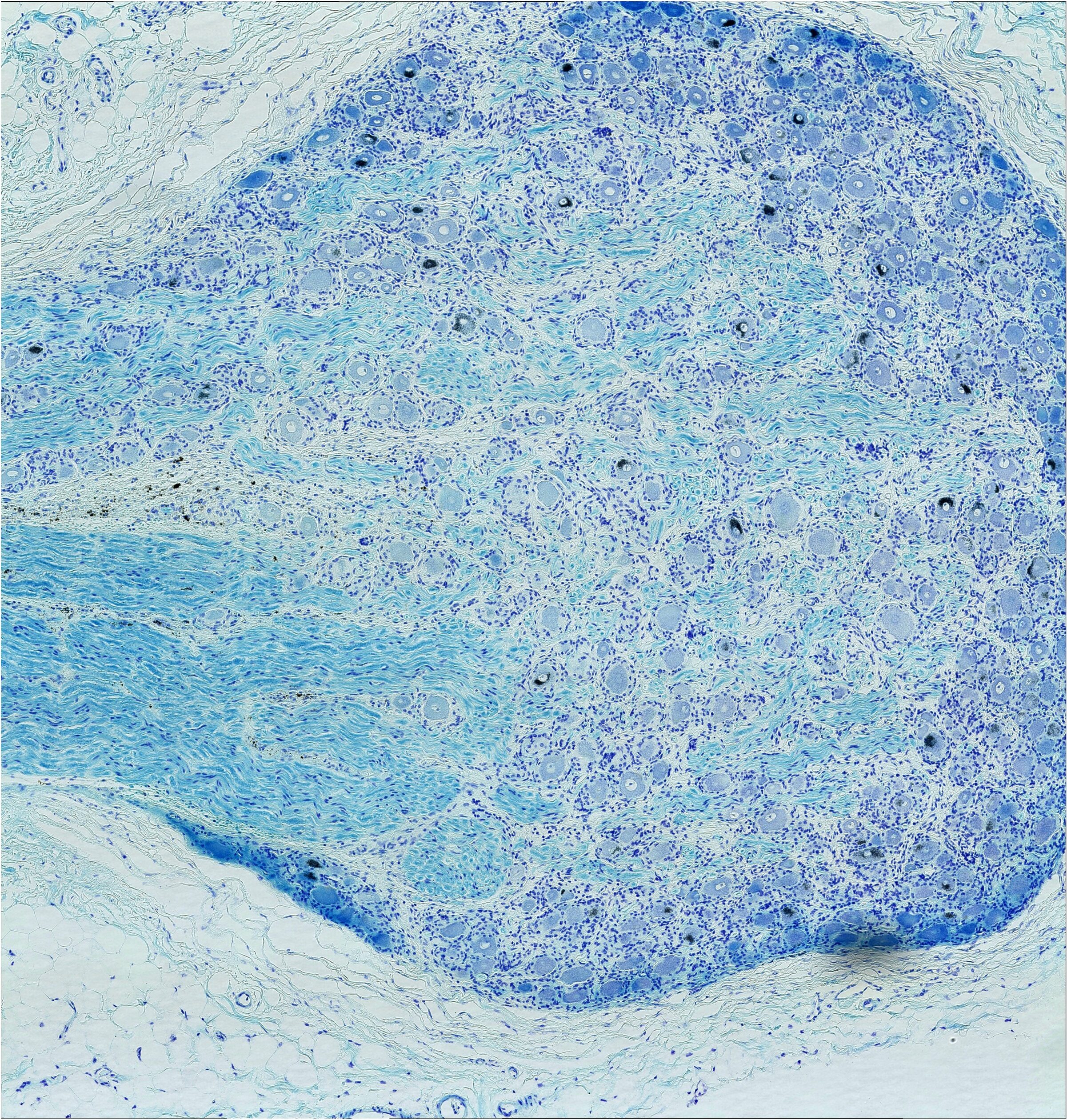
IMAGE 1 – Luxol fast blue staining with hematoxylin counterstaining at 2X magnification. Image shows myelinated fibers exiting the human dorsal root (upper left part of image). (Credit: Michael Iadarola, Matthew Sapio)
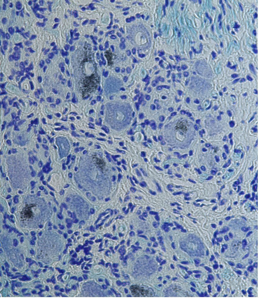
IMAGE 2 – Image taken at 40X magnification of the same DRG section. Satellite cells tightly arranged around neuronal cell bodies. (Credit: Michael Iadarola, Matthew Sapio)
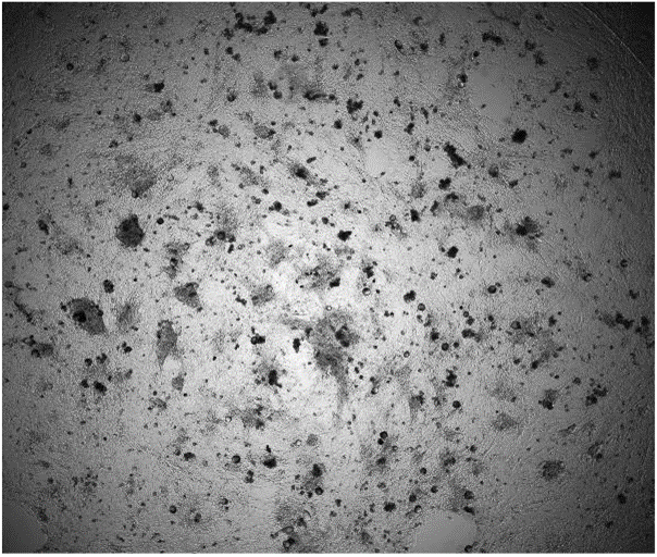
IMAGE 3 – Wide field image at 4X magnification of human dorsal root ganglia neuronal culture at 9 days in vitro (DIV). The neuronal culture consists of DRG neurons and non-neuronal cells. At DIV 9, the culture is confluent and neurites have re-grown on the neurons (see immunostained image) and the non-neuronal cells have spread and grown. (Credit: Yannick Miron, Fanny Chen, AnaBios)
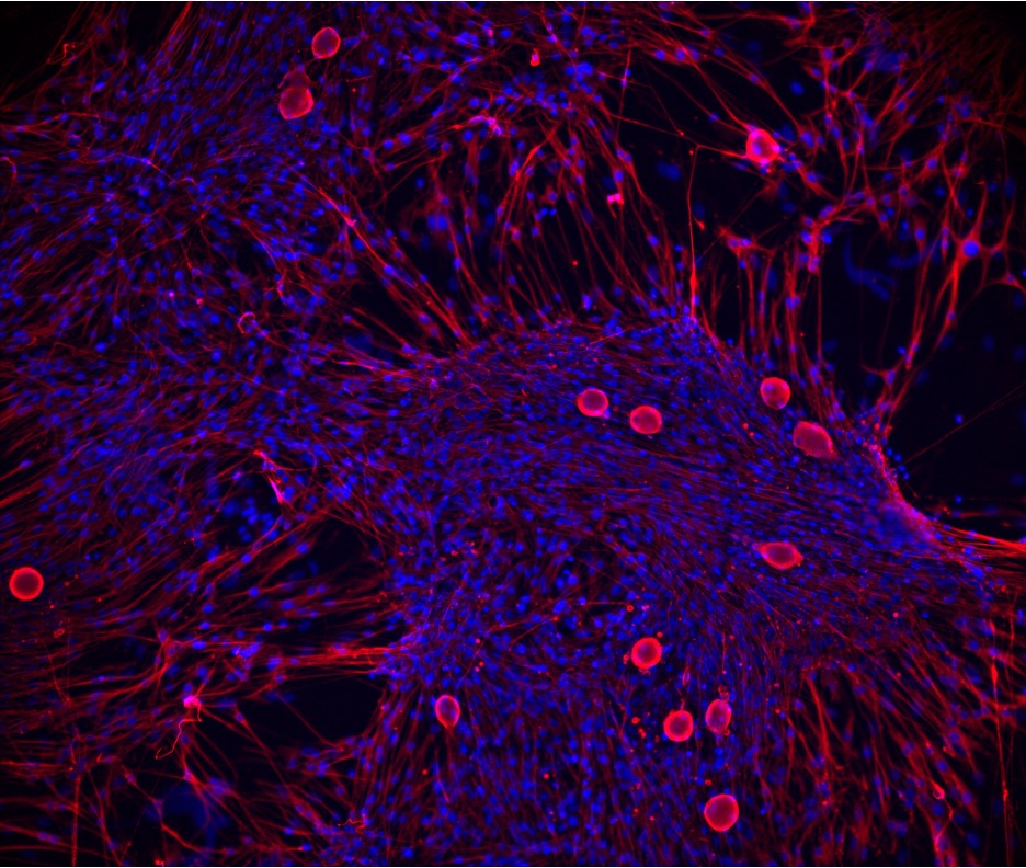
IMAGE 4 – Fluorescent image of immunostained fixed human dorsal root ganglia neuronal culture take at DIV 15. Red is beta-III-tubulin (neuronal marker) and blue is Hoescht dye (cell nuclei). Production of live human DRG neuronal cultures is described in Davidson et al (2014) Pain. Human sensory neuronal culture (Credit Tiara Wang, AnaBios)
Alpenglow Biosciences recently created new video of human DRG samples from AnaBios using their unique version of light sheet microscopy.
Staining: computation H&E (nuclei: ToPro3, protein: Eosin)
Microscope Meta-Data
Zoom image: 638 nm Excitation – To-PRO-3; 561 nm excitation – Eosin; 41.3 GB raw data; 0.3 x 0.25 x 0.25 mm X,Y,Z dimensions; Scan time: 7 minutes, 5 Seconds
PAIN RESEARCH WEBINARS SPONSORED BY ANABIOS


HAVE MORE QUESTIONS?
CONTACT US
To inquire about products, services and pricing, please go to the ‘Contact Us’ page by clicking the button below.